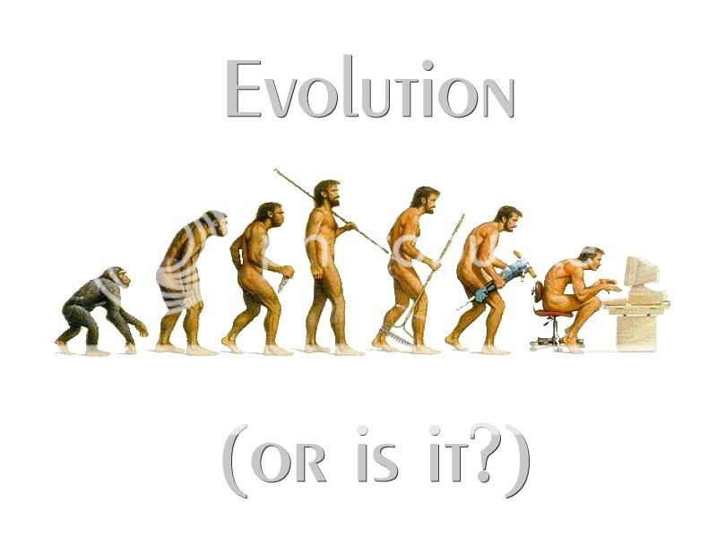
Wings on a flightless bird, eyes on a blind fish, and sexual organs on a flower that reproduces asexually—the casual observer might ask, what’s the point? But these vestigial organs and structures, once useful in an ancestor and now diminished in size, complexity, and/or utility, carry important information and give us clues to our evolutionary past.
Though humans often think of vestigial organs as useless little fixtures that sometimes, as in the case of the appendix, cause us extreme anguish, we wouldn’t know nearly as much about macroevolution as we do now without their presence. In On the Origin of Species, Charles Darwin used vestigial organs as evidence for evolution, and their presence has helped define and shape our phylogenetic trees.

Why the Leftovers?
Contrary to what most think, vestigial doesn’t necessarily mean useless; in some cases, we may just not yet know exactly how the organ is used in its current incarnation. (The human thymus was once thought to be vestigial). Because these structures can be traced back through the ancestors, they essentially serve as a marker of evolution; no organism can have a vestigial organ that hasn’t been found in its forefathers. For this reason, you won’t ever find feathers on a mammal or gills on a primate.
Similar in concept to vestigial structures are atavisms, which are the reappearance of a structure or trait that isn’t found in the immediate ancestors. For instance, whales and dolphins have been found in nature with hind limbs; this rare occurrence is due to the reemergence of a trait they inherited from their terrestrial ancestors.
Humans also contain structures that mark where we came from and perhaps, which structures’ evolution will take care of over time.
Human Tail (Bone)

One striking example of an atavism is the human coccyx, or tailbone, which is a relic of the mammalian tail. Useful for mammals that use tails for balance, species-to-species signaling, and support, the tail is missing in apes and in humans. However, all human embryos initially have a tail. Normally, they regress into four to five fused vertebrae (the coccyx). However, there have been numerous case studies of human children being born with an extended coccyx—a tail—that was removed without incident. Ranging from one inch to five, the gene that normally stops vertebrae elongation is decreased and the human tail remains at birth.
Wisdom Teeth

Our ancestors, known to be herbivores, needed strong molars for mashing up and chewing plant material. This relic is why many of us will develop wisdom teeth, also known as third molars. Theoretically, they could still be used for chewing, but in one third of people, they can come in sideways, impacted, or can cause pain and infection. This is why these vestigial structures are almost always removed when they begin to come in.
Appendix

Another leftover from our plant eating ancestors is the vermiform appendix, which is an organ attached to the large intestine. A similar sac is much bigger in other animals than it is in humans and is used to aid in digesting high cellulose diets.
While appendicitis can be a potentially fatal condition, and removing the appendix has no adverse effects, some researchers think that the appendix might have an auxiliary function, such as aiding the immune system.
Vitamin C Synthesis

In humans, vitamin C deficiency causes scurvy, and can eventually cause death. We can’t synthesize vitamin C (ascorbic acid), but our ancestors, save for the guinea pig and primates, were able to do so. Therefore, it makes sense that we have a vestigial molecular structure, now defunct, that manufactures the vitamin. The gene required for vitamin C synthesis was found in humans in 1994, but it was a pseudogene, meaning it was present but unable to function. The pseudogene was also found in some primates and guinea pigs, as expected.
Male Nipples

Male nipples are sometimes referred to as vestigial, although they aren’t truly, because they were never functional in our ancestors. Instead, they most likely occur because in the embryonic stage we are essentially sexless, only differentiating into male and female with the presence of hormones.
Goose Bumps

When we get goose bumps, it’s the action of muscle fibers called erector pili, which cause the hairs in follicles to stand to attention. In animals, such as a cat, this causes a larger appearance and can be used to thwart an attacker, as well as trap air between feathers and fur for insulation. However, humans, with our minimal coating of fur, don’t really need the raised hair; we use jackets instead. It is therefore thought that goose bumps don’t really serve much of a purpose. However, the small expenditure of energy used to contract the muscles could, perhaps, cause a tiny release of heat. Or, because goose bumps are associated not only with cold, but emotional responses as well (listening to a good song, seeing a scary movie) they could now serve as a form of communication with others.
Vomeronasal Organs (VNOs)

In mice and other animals, the tiny vomeronasal organs (VNOs) are thought to be responsible for pheromone detection, helping to pick up the chemicals that signal a potential mate, reproductive status, and other social cues. Although similar structures have been found in humans, they’re largely thought to be vestigial and inactive, having lost nerve connection to the brain.
There are other vestigial and atavistic structures in humans, especially when you consider the potential leftovers in our genomes. And if they don’t require too much energy or resources to make, chances are they’ll stick with us for the long haul.
National Institutes of Health, History of Medicine.
---
More organs we can live without
Our bodies are miraculous machines comprised of bones, cartilage, arteries, veins, nerves, tissue and organs. Everything works together to keep us in optimal health.Yet, like machines, sometimes our bodies break down. If an organ fails, removal might be an option.
Here are some of the organs that we can give up:
Uterus

Function: The uterus or womb is the organ where the fetus matures during pregnancy. It is present when a female is born but not mature until puberty, when menstruation begins. Ovaries are independent organs with their own blood supply.
Why remove it: The most common diseases of the uterus are fibroids, benign muscle growths within the uterus wall; endometriosis, when endometrial tissue grows outside the uterus causing bleeding and pain; and endometrial cancer, according to Dr. Gerald Harkins, medical director of minimally invasive gynecological surgery at Penn State Milton S. Hershey Medical Center. Removal of the uterus is called a hysterectomy, which is one of the most common surgeries in the U.S. today. “Most hysterectomies are done laproscopically, with the option to keep the cervix,” Harkins says. “Removal of the uterus does not necessarily include removal of the ovaries.”
Living without it: Without a uterus, a woman cannot physically deliver a child nor will she menstruate. However, women who have had a hysterectomy but whose ovaries have not been removed and who desire children can donate their eggs to a surrogate. Removal of the uterus doesn’t affect a woman’s hormonal status if her ovaries are not removed. The American College of Obstetrics and Gynecology recommends that women up to age 65 keep their ovaries at surgery, and research shows that women with ovaries have improved longevity.
Kidney

Function: Kidneys are bean-shaped paired organs, located in the retroperitoneal space with one on each side of the spine. Most people only think of kidneys as organs that clear toxins from the body and produce urine. However, they also produce a hormone that allows the body to make red blood cells. According to Dr. Jennifer Kogan of the Hospital of the University of Pennsylvania, the kidneys also help to maintain electrolytes (sodium, potassium, calcium) and water balance. They are important in bone and vitamin D metabolism and play a role in regulating blood pressure.
Why remove it: The most common cause for removing a kidney, known as a nephrectomy, is to remove a kidney mass when there is concern for cancer, according to Dr. Harold C. Yang of the PinnacleHealth Transplantation Services. A kidney may also be removed when there is a congenital malformation causing recurrent kidney infections. Kidney donation is another common cause for removal.
Living without it: Everyone has two kidneys, although only one kidney is actually needed. However, it is important that individuals who have had a nephrectomy take special precautions to protect their one existing kidney by avoiding medications that can injure their remaining kidney and by eating healthy and avoiding alcohol
Gallbladder

Function: The gallbladder is the hollow storage organ for bile made in the liver. When it’s healthy, it stores and concentrates some bile and squeezes it into the small intestines where it helps aid digestion.
Why remove it: According to Kunkel, when substances in the bile crystallize, they become solid and are referred to as gallstones. In some instances, gallstones cause no symptoms. In others, they can irritate the wall of the gallbladder or cause blockage of the nearby ducts which, in turn, causes inflammation of the gallbladder, pain, nausea and infection.
Most commonly, a person eats a fatty meal, sending the gallbladder into overtime, which may force a gallstone to block a duct. As the gallbladder tries to contract, the patient gets serious cramps and abdominal pain. Gallbladder removal is often elective, based on seriousness and worsening of symptoms. However, if the pain is unrelenting, the patient has a fever and elevated blood count and an ultrasound shows inflammation, surgery should be immediate to prevent perforation. Often the surgery is four tiny incisions done laproscopically, which takes 45 minutes to one hour. A doctor may follow up with an imaging study to make sure no stones have gotten into the main bile duct. Often the patient can return home the same day, with some soreness for up to five days.
Living without it: When a gallbladder is removed, the liver learns how to compensate by pushing the bile directly into the intestines. It is important to eat a balanced diet, high in fiber.
Spleen

Function: The spleen, located in the upper left quadrant of the abdominal cavity, is the filter for aged red blood cells when they are out of commission and can no longer carry oxygen. It is also part of the lymphatic system, which fights infection and keeps body fluids in balance. Recent researchers have found it is a reservoir for huge numbers of immune cells call monocytes. In the event of a serious illness (heart attack, gashing wounds, microbial invasion), the spleen will send these monocytes multitudes into the bloodstreams to help with the crisis.
Why remove it: According to Kunkel, a spleen is most often removed when it becomes diseased (cancer or immunological disease), which will cause it to become enlarged and painful. In addition, a spleen can also be ruptured, often the result of an injury in a car or motorcycle accident, and will need to be removed. Kunkel often removes the spleen laproscopically, depending on the condition of the organ.
Living without it: Without a spleen, other organs such as your liver, will take over some of the spleen’s work. However, the spleen definitely has an immunological component to fight infections, and thus, vaccines are given to help fight off infection potential. If possible, it is best to administer the vaccines before surgery.


No comments:
Post a Comment